
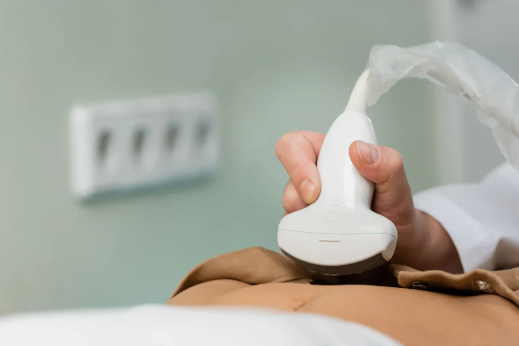
Ultrasound Examination of the Liver, Gallbladder, Bile Ducts, Pancreas, Spleen and Bowel
This is a non-invasive imaging examination that uses high-frequency sound waves to
produce images of the organs within the abdominal cavity.
- Liver: The liver is examined for the diagnosis of conditions such as fatty liver disease (hepatic steatosis), hepatitis, cysts, tumors and cirrhosis.
- Gallbladder and Bile Ducts: The gallbladder and bile ducts are examined for the detection of gallstones, inflammation (cholecystitis) and bile duct obstruction.
- Pancreas: The pancreas is examined for the detection of conditions such as pancreatitis, cysts or tumors.
- Spleen: The spleen is examined for abnormalities in size (e.g. splenomegaly), injuries or tumors.
- Bowel: The bowel is examined to exclude bowel obstruction (ileus), appendicitis and inflammatory bowel diseases such as Crohn’s disease and ulcerative colitis.
Patients are usually advised to fast for 6–8 hours before the examination to allow better visualization of the abdominal organs.
You may also be asked to drink water before the examination, as this may help improve the visualization of certain organs.
During the ultrasound examination, the patient lies on an examination table and the doctor applies a gel to the abdominal area to ensure optimal transmission of the ultrasound waves.
A transducer (ultrasound probe) is then moved over the abdomen to obtain images of the internal organs.
The results of the ultrasound examination can help diagnose a wide range of conditions and guide further treatment.
Abdominal ultrasound is a fundamental and often first-line imaging examination for the investigation of symptoms such as abdominal pain, jaundice and unexplained weight loss.


